RRV
Rose rosette virus (RRV) causing rose rosette disease (RRD) produces symptoms of excessive thorns and red pigment, dense shoot growth, and eventual death in roses. Eriophyid mites and grafting spread RRV. There are no cures for infected plants which should be destroyed.
History
Rose rosette disease (RRD) was first recorded in 1940-1941 in Manitoba Canada, and California, Nebraska, and Wyoming USA causing symptoms of excessive shoots, thorns, and red pigmentation on roses (3). This disease was of particular interest for use as a potential biocontrol agent for the invasive multiflora rose. Multiflora roses were promoted for use from the 1930’s-1970’s as an attractive living fence to control soil erosion and provide food for animals (rose hips and leaves) (7). Multiflora roses were quite hardy and prolifically reproduced by both seed, crown growth, and from branches that bent over and rooted severely reducing pasture and park quality. Physical removal, herbicides, and overgrazing by goats are expensive control measures so a method that would kill the plants without requiring a lot of physical labor or chemicals was desired (7).
RRD was studied for its use as a biocontrol of multiflora roses and was found to kill multiflora roses in 2-3 years for single crowned plants and 4-5 years for multicrowned plants (7). RRD was found to infect only wood’s rose, multiflora rose, macartney rose, hybrid tea rose (and all other cultivated roses including shrub, floribunda, grandiflora, miniature), and sweetbriar and was recommended for use to control multiflora roses (7). This may be surprising because of the high popularity of cultivated roses today, however it is only with the release of the Knock Out® shrub rose line in 2000 that consumers embraced the floriforous, disease resistant, cold hardy, heat resistant new generation of roses buying 4 million Knock Out® roses a year for the next four years (10) (followed by many more shrub roses as new varieties were released). Prior to the Knock Out® shrub rose line the majority of cultivated roses required pesticide sprays to control various pathogens, special pruning, and protection to survive winter.
The organism causing RRD was not described until 2011 when Rose rosette virus had the genome sequenced and in 2015 the experiments were completed to show it caused RRD (8, 6). RRV can be found throughout the USA, Canada, and was recently identified in India (3, 9, 4).
Transmission
RRV has been shown to be transmitted by the eriophyid mite, Phyllocoptes fructiphilus, and through grafting (1, 2). This species of mite is 140-170 micrometers (0.0055-0.0067 inches) long by 43 micrometers (0.0017 inches) wide and is found in rose shoots and buds so is difficult to observe without magnification (2). These tiny mites are thought to be passively moved by air currents or humans (2).
Studies conducted in the field in West Virginia revealed that plants infected by eriophyid mites carrying RRV took from 30-279 days to express symptoms (1, 2).
Symptoms
Early RRV symptoms include mosaic leaf patterns. Later symptoms include excessive thorns on canes, high levels of red pigmentation in canes and leaves, excessive lateral shoot growth, and dense masses of shoots (witches’ brooming). RRV infections will kill infected plants in 2-5 years (7).
 A Knock Out® rose with healthy growth on the left and RRV infected growth showing excess red pigmentation and witches’ brooming of a flower shoot.
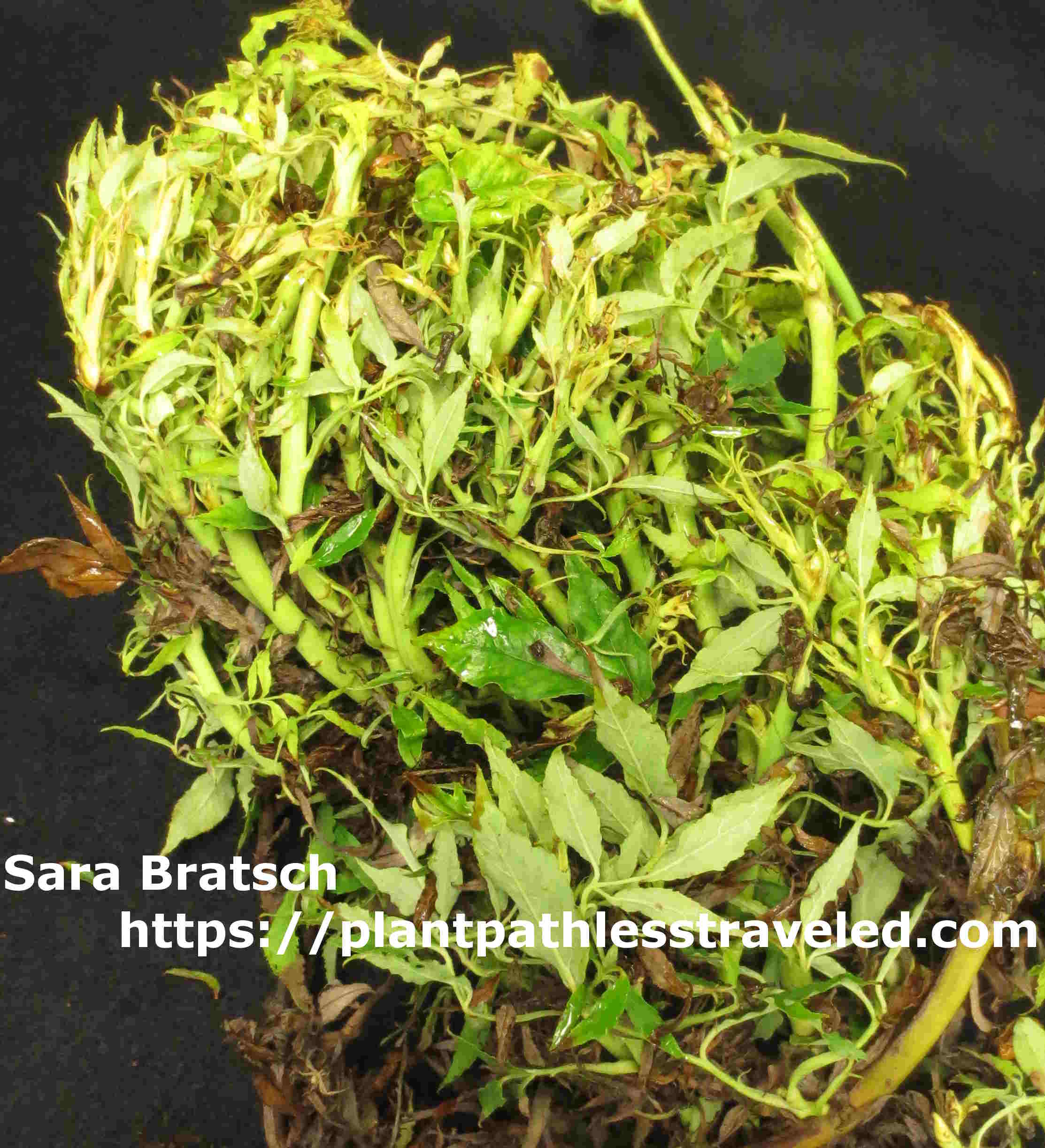 A “White Smith Perish” rose infected with RRV showing excessive lateral shoot growth and witches’ brooming.
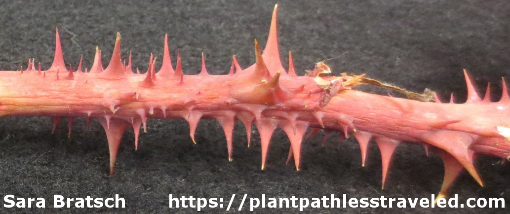 A Knock Out® rose cane with leaves removed to show excessive thorniness and red pigmentation.
Not symptoms: new growth
Many varieties of roses have been released that have very dark red or purple new growth that fades to green within a few weeks. If the rose variety is unknown check for the presence of excessive thorns among the reddened foliage. RRV infected roses with reddened tissue will not fade in color.
 A Knock Out® rose with new red growth. This plant is healthy and RRV negative.
Not symptoms: herbicide exposure
Accidental herbicide exposure can cause distorted, RRD-like symptoms that will disappear with later growth. If there are any other plants that have odd growth it's worth investigating if a neighbor, lawn care company, or city/county sprayers were out spraying chemicals. Symptomatic tissue can be submitted to an herbicide testing lab to get confirmation that herbicide exposure caused the symptoms.
Diagnosis
Current testing methods to diagnose RRV are based on amplifying a portion of the virus genome by reverse transcription polymerase chain reaction (RT-PCR) (5). This molecular test can be conducted by a plant disease clinic or diagnostic lab. Work is on-going to produce a rapid, protein based method for use in the field or garden.
Management
Large and valuable rose collections should be observed frequently for symptoms of RRD. Plants showing symptoms can be submitted to a plant disease clinic or diagnostic lab for testing. There is no cure for a plant virus infection and the plant must be destroyed. Infected plants should be bagged (to prevent infected mites from escaping) and removed (including roots) from the garden. Pruning symptomatic portions out of a bush does not cure it as the plant is infected systemically (from the roots to the leaves). Any neighboring roses should be watched for the development of symptoms and removed.
Work is currently ongoing to determine if any cultivated rose varieties are resistant to RRV. When choosing new rose plants for your garden chose roses that have tested negative for RRV or have no symptoms.
All photos taken by Sara Bratsch. For non commercial use only.
Please contact regarding all other uses including but not limited to: data for computational algorithms, books, pamphlets, articles, apps, websites, etc.
Cite this article:
Bratsch, Sara. 2017. "RRV". Web article. (date accessed). http://plantpathlesstraveled.com/RRV/
Citations
- Allington, W. B., R. Staples and G. Viehmeyer. 1968. Transmission of rose rosette virus by the eriophyid mite Phyllocoptes fructiphilus. J. Econ. Entomol. 61:1137-1140.
- Amrine Jr, J. W., Hindal, D. F., Stasny, T. A., Williams, R. L., & Coffman, C. C. (1988). Transmission of the rose rosette disease agent to Rosa multiflora by Phyllocoptes fructiphilus (Acari: Eriophyidae). Entomological news (USA). 99:239-252.
- Conners, L. 1941. Twentieth Annual Report of the Canadian Plant Report Survey, 1940. 98.
- Chakraborty, P., Das, S., Saha, B., Karmakar, A., Saha, D., & Saha, A. (2017). Rose rosette virus: An emerging pathogen of garden roses in India. Australasian Plant Pathology, 46(3), 223-226.
- Dobhal, S., Olson, J. D., Arif, M., Suarez, J. A. G., and Ochoa-Corona, F. M. 2016. A simplified strategy for sensitive detection of Rose rosette virus compatible with three RT-PCR chemistries. J.Virol. Methods.232:47-56.
- Di Bello, P. L., Ho, T., & Tzanetakis, I. E. (2015). The evolution of emaraviruses is becoming more complex: seven segments identified in the causal agent of Rose rosette disease. Virus research, 210, 241-244.
- Epstein, A. H., & Hill, J. H. (1998). Status of rose rosette disease as a biological control for multiflora rose. Plant disease, 83(2), 92-101.
- Laney, A.G., Keller, K.E., Martin, R.R. and Tzanetakis, I.E. 2011. A discovery 70 years in the making: characterization of the Rose rosette virus. J. Gen. Virol. 92:1727-1732.
- Martin, C.W. 2014. Rose Rosette Disease and the Impacts on Propagation©. Acta Hortic. 1055:319-321.
- Pemberton, H. B., and Karlik, J. F.2013. A Recent history of changing trends in USA garden rose plant sales, types, and production methods.VI International Symposium on Rose Research and Cultivation. 1064:223-234.